
Multiwavelength LED Source with a Single Fiber Output
A Scientific Multiwavelength LED Source is constructed using high-brightness LEDs that are optically combined into a single fiber or a single lightguide. Customers can select from a wide range of LED wavelengths, spanning from Deep UV, UV, Visible, to NIR and SWIR, to meet their specific requirements.
High Power Fiberoptic LED Light Source
High-power fiber-coupled LED light sources are available across a broad spectrum of wavelengths, spanning from UV to NIR and SWIR. These versatile and favored scientific light sources find applications in various fields. Power control is conveniently achieved using a 10-turn dial, while computer control can be accomplished via TTL and analog inputs or an optional USB link. The unparalleled convenience of these light sources becomes even more pronounced when used with flexible, unbreakable polymer optical fibers (POF).

Multi-Channel Fiber-Coupled LED Source
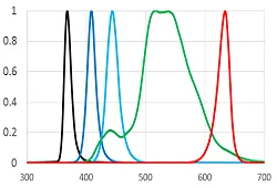
The Multiwavelength Fiberoptic LED source is a cutting-edge device that offers two or more High Power LED sources in a single unit. Each channel of this multi-channel LED source features an independent high current driver with TTL and Analog Input control, providing maximum flexibility and precision in operation. In addition, computer control is also possible via USB, making it easy to integrate into experimental setups.
High Brightness Fiber-Coupled White Light Source
Prizmatix High Brightness Fiber-Coupled White Light Source is a remarkable tool for generating powerful, incoherent, Cold White light from thin optical fibers. This innovative light source utilizes the KYOCERA SLD Laser component source, which uses a high-power laser to illuminate a phosphor, resulting in a bright, reliable, and efficient white light source.
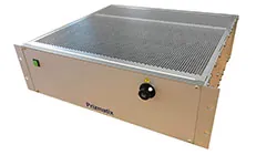
Ultra High Power Fiber-Coupled LEDs for Scientific Research
The product line uses the brightest and most powerful LED chips to create most powerful fiberoptic LED light sources at various wavelengths for numerous power-hungry scientific applications. This scientific LED source designed as a fully integrated module, including the LED high current driver, control electronics, thermal management making it a versatile and adaptable laboratory light source for a wide range of scientific and industrial applications.

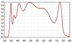
Fiber Coupled Broadband White LEDs for Spectroscopy
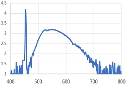
The FC-BBW50-Plus is a high-quality fiber optic broadband White LED light source that delivers exceptional performance. This advanced light source features a high CRI, SunLike white LED along with additional UV-Violet and Near IR LEDs, which combine to produce a wide emission spectrum ranging from 390nm to 750nm. This fiber coupled broadband White light source is ideally suited for a range of reflection and transmission spectroscopy applications. With its self-contained design, this Scientific LED source includes all necessary driver electronics and thermal management, eliminating the need for an external controller box. Simply plug in the included DC power adaptor and you're ready to go.
© |